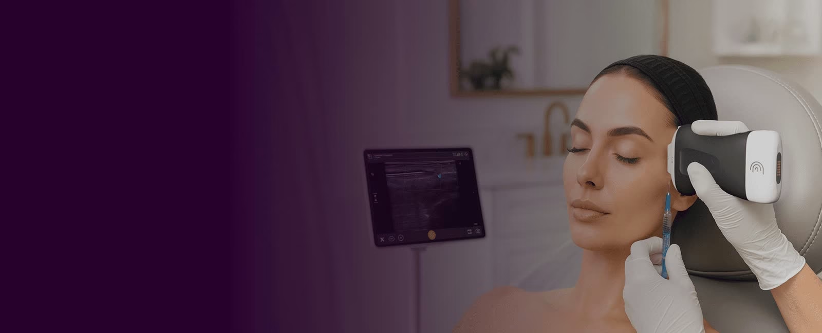
Surface examination shows only part of the picture. Clarius is a wireless, high-frequency ultrasound scanner that connects to a compatible smart device and lets a trained provider view superficial anatomy in real time when that information can help answer a clinical question.
In ultrasound-guided aesthetics, the provider uses a handheld ultrasound scanner to look beneath the skin before, during, or after selected procedures.
Depending on the case, a trained provider may review facial tissue planes, map relevant vessels with Doppler, assess prior filler, or visualize a needle or cannula during a procedure.
Ultrasound adds information; it does not eliminate procedural risk or replace training, anatomy knowledge, sterile technique, informed consent, or clinical judgment.
Ultrasound is not a step promised with every injectable. Your provider decides whether imaging is useful, what it shows, and how findings affect your plan. Only trained, authorized providers scan or perform ultrasound-guided procedures under AestheteMed protocols.

Clarius is used selectively, based on provider assessment, training, the treatment area, and the clinical question.
Visualize the location and depth of vessels to support individualized treatment planning.
Review tissue planes and key anatomy before selected filler or biostimulator procedures.
Support visualization of a needle or cannula when the provider determines guidance is appropriate.
Help locate and characterize previous filler when relevant to the treatment plan.
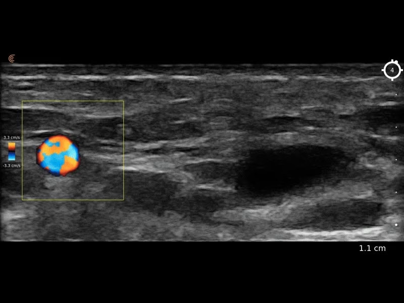
Use Doppler imaging to add vascular information during selected evaluations.
Add real-time imaging to evaluation and management when clinically indicated.
Sample Doppler ultrasound image for illustration. Not a patient finding.
Imaging adds a view of anatomy that is unavailable from surface examination alone.
Review relevant vessels, soft tissue, and prior filler before treatment planning.
Support visualization of instruments and tissue planes when guidance is clinically useful.
Help evaluate selected concerns or locate existing filler.
Imaging happens at the treatment chair during your visit.
Visualize blood flow and vessels in selected areas.
Findings are used together with history, examination, and clinical judgment.
How ultrasound fits into your visit.
Imaging is used when the provider determines it can help answer a clinical question.
Only trained, authorized providers perform scans and ultrasound-guided procedures.
Ask during your consultation whether ultrasound is planned for your treatment.
Simple steps. Better context.
History, goals, previous treatments, and physical examination.
Identify the anatomy relevant to the clinical question.
Use imaging findings to support the provider's procedure plan.
Save useful images and findings within the approved clinic workflow.
Answers to common questions about ultrasound in aesthetic treatments.
No. Ultrasound is used selectively when the provider determines imaging is clinically useful.
Depending on the area, it can show skin layers, soft tissue, vessels and blood flow with Doppler, and existing filler.
No. Imaging adds information but does not eliminate procedural risk or replace training and clinical judgment.
No. The handheld probe is placed on the skin with gel; it is non-invasive.
In selected cases it can help locate and characterize previous filler when relevant to your treatment plan.
Aesthetemed is a med spa and wellness clinic in Hallandale Beach, FL, serving patients from Miami, Fort Lauderdale, Aventura, Hollywood, and Sunny Isles Beach. All medical treatments are performed under medical supervision.
Every patient receives a personalized consultation and a treatment plan built around their goals. AM Club members get preferred pricing across clinic services.

Injectable treatments where imaging may be useful.
Message AestheteMed on WhatsApp to ask whether ultrasound is planned for your treatment.
By submitting this form, you agree to receive emails about our products, sales, exclusive content and more. View our Privacy Policy.
© 2025 AestheteMed. All rights reserved.